Rapid Nanofiber Spinning Fills the Gap in Small-Diameter Vascular Grafts
Key Takeaways
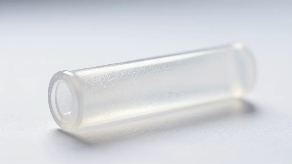
- •FRJS creates 0.5 mm diameter grafts in under 90 seconds.
- •Grafts withstand 276 mmHg pressure, exceeding normal blood pressure.
- •Endothelial layer formed within one week; smooth muscle infiltrated in two weeks.
- •No thrombosis observed in 12 rats over four weeks.
- •Sterilization and solvent removal fit within intra‑operative time frame.
Pulse Analysis
Small‑diameter vascular grafts have long been a blind spot in cardiovascular surgery. Conventional synthetic tubes such as expanded PTFE work well above 6 mm but fail catastrophically when scaled down, forcing surgeons to harvest autologous veins—a process that adds operative time, creates a secondary wound, and limits graft availability. Tissue‑engineered alternatives promise biocompatibility but require weeks of cell culture, making them impractical for emergency repairs. The market therefore demands a rapid, scalable manufacturing method that delivers nanoscale cues essential for cell organization while matching patient‑specific geometry.
Focused rotary jet spinning (FRJS) bridges that gap by marrying high‑throughput fiber production with shape‑controlled deposition. A polymer solution—here poly(L‑lactide‑co‑ε‑caprolactone) (PLCL)—is expelled through a high‑speed rotor, forming nanofibers that are guided by a focused air stream onto a rotating mandrel. By adjusting mandrel orientation, fiber alignment and wall thickness can be tuned independently, enabling seamless tubes, bifurcations, or curved conduits. Production times drop from hours (electrospinning) to under ten minutes, and mechanical testing shows the grafts tolerate pressures well above normal systolic levels (276 mmHg) and provide superior suture retention, addressing two critical failure modes of existing prostheses.
Preclinical data reinforce the technology’s promise. In a rat femoral model, both arterial and venous grafts maintained patency, oxygen saturation, and hemoglobin for four weeks, with no evidence of thrombosis. Endothelial cells formed a confluent lining within a week, while induced‑pluripotent‑stem‑cell‑derived smooth‑muscle cells infiltrated the wall by two weeks, indicating early remodeling. Sterilization via hydrogen‑peroxide plasma and rapid solvent removal fit within a typical surgical window, suggesting intra‑operative deployment is feasible. Continued studies in larger animal models and scaling of polymer options will be essential to align degradation rates with tissue regeneration, but FRJS positions itself as a disruptive platform poised to fill a critical unmet need in vascular surgery.
Rapid nanofiber spinning fills the gap in small-diameter vascular grafts
Comments
Want to join the conversation?