Capturing the Moment of Organelle Handoff Inside Living Cells
•January 28, 2026
0
Why It Matters
The discovery links ER remodeling to intracellular cargo transport, offering new targets for diseases involving autophagy or cytoskeletal dysfunction. It also establishes a powerful imaging paradigm for real‑time, label‑free cellular biology.
Key Takeaways
- •Real‑time visualization of autophagosome transfer from ER to microtubules
- •ER three‑way junctions act as organelle handoff hubs
- •DySLIM enables millisecond temporal, nanometer spatial resolution
- •Disrupting microtubules halts long‑range organelle transport
Pulse Analysis
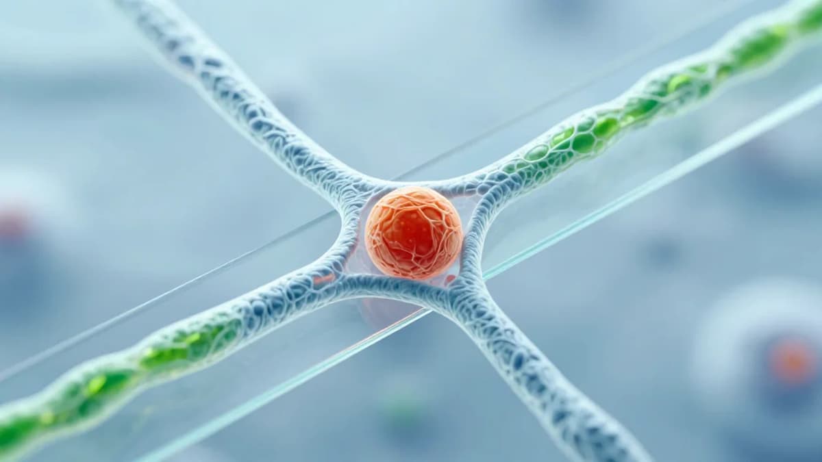
The endoplasmic reticulum (ER) has long been viewed as a static scaffold for protein synthesis, but recent evidence suggests it actively orchestrates intracellular logistics. Autophagosomes—double‑membrane vesicles that sequester damaged proteins—must exit the ER and hitch a ride on microtubule highways to reach lysosomes. This limitation has hindered quantitative models of cargo distribution across the cytoplasm.
The breakthrough came from merging interferometric scattering microscopy with fluorescence tagging in a custom DySLIM platform. By labeling LC3, the team visualized autophagosomes and microtubules simultaneously, while DySLIM provided label‑free, nanoscopic maps of the ER network at millisecond intervals. This dual‑mode approach captured organelles drifting along ER tubules, pausing at three‑way junctions, and then launching onto adjacent microtubules with nanometer precision—offering the first direct, real‑time view of organelle handoff portals. The system also recorded ER tubule extension as organelles pulled the network forward, linking cargo movement to membrane remodeling.
Beyond satisfying a basic cell‑biology curiosity, these observations reshape our understanding of how organelle traffic influences ER morphology and vice versa. The dependence on intact microtubules for long‑range transport highlights potential therapeutic targets in diseases where autophagy or cytoskeletal integrity is compromised. Moreover, the DySLIM methodology sets a new benchmark for label‑free, high‑speed nanoscopy, promising broader applications in drug screening, neurodegeneration research, and the development of next‑generation imaging platforms. Future integration with cryo‑electron tomography could bridge the gap between live‑cell dynamics and atomic‑level structural insight.
Capturing the moment of organelle handoff inside living cells
0
Comments
Want to join the conversation?
Loading comments...