Corneal Nerve Regeneration via MSC‐Derived EVs: Tissue Source and Culture Dimensionality Dictate miRNA Cargo and Therapeutic Efficacy (Small 6/2026)
•January 27, 2026
0
Companies Mentioned
Why It Matters
The study proves that 3D‑engineered MSC‑EVs can be mass‑produced with consistent potency, accelerating cell‑free therapies for corneal nerve damage and broader ocular regeneration.
Key Takeaways
- •3D MSC culture triples EV yield.
- •3D EVs carry enriched neurotrophic miRNAs.
- •Tissue origin alters EV miRNA profile.
- •3D‑derived EVs restore corneal nerves in vivo.
- •Framework guides cell‑free ocular therapy design.
Pulse Analysis
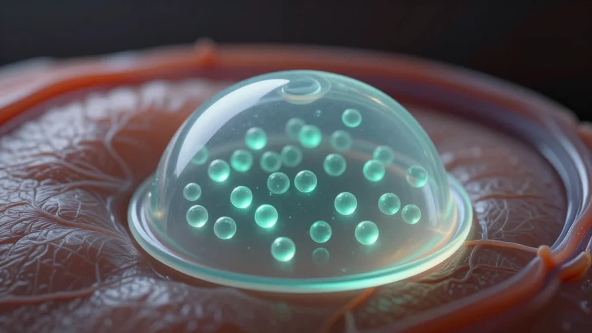
Extracellular vesicles (EVs) have emerged as a versatile platform for delivering bioactive molecules, especially microRNAs, to damaged tissues. In regenerative ophthalmology, the delicate architecture of the cornea and its dense innervation demand highly targeted interventions that avoid the risks associated with cell transplantation. However, EV therapeutic performance is highly variable, depending on the parent mesenchymal stem cell (MSC) source and the conditions under which those cells are expanded. Understanding how tissue origin and culture dimensionality shape EV composition is therefore critical for translating cell‑free strategies from bench to clinic.
The Small study demonstrates that culturing MSCs in three‑dimensional (3D) scaffolds not only boosts vesicle output but also reprograms the miRNA cargo toward a neurotrophic signature. Compared with conventional two‑dimensional plates, 3D‑grown cells produced roughly three times more EVs, with a marked increase in miR‑21, miR‑124, and miR‑146a—miRNAs known to promote axonal growth and extracellular matrix remodeling. This enrichment translates into superior functional outcomes: animal models of corneal injury treated with 3D‑derived EVs showed faster nerve re‑innervation and restored corneal sensitivity.
These findings lay a practical blueprint for scalable, cell‑free ocular therapies. By selecting an optimal tissue source and leveraging 3D bioprocessing, manufacturers can generate standardized EV batches with predictable potency, addressing a key regulatory hurdle for biologics. The approach also opens avenues for engineering EVs with bespoke miRNA panels to treat a broader range of neuro‑ophthalmic disorders, such as neurotrophic keratitis or diabetic retinopathy. As the market for regenerative eye treatments expands, 3D‑enhanced MSC‑EVs could become a cornerstone of next‑generation precision medicine.
Corneal Nerve Regeneration via MSC‐Derived EVs: Tissue Source and Culture Dimensionality Dictate miRNA Cargo and Therapeutic Efficacy (Small 6/2026)
0
Comments
Want to join the conversation?
Loading comments...