From Slices to Whole Bodies: How 3D Cell Atlases Could Reshape Pathology Research
Why It Matters
The ability to visualize and quantify cell distributions across entire organs transforms disease modeling, drug safety assessment, and the future of 3D pathological diagnosis, offering unprecedented precision for biomedical research and clinical translation.
Key Takeaways
- •CUBIC method cleared entire neonatal mouse for imaging.
- •3D atlas maps every cell across organs and whole body.
- •Enables comparison of cell distributions across experimental conditions.
- •Integrates with spatial transcriptomics for molecular‑morphology insights.
- •Paves way for next‑gen 3D pathological diagnostics.
Pulse Analysis
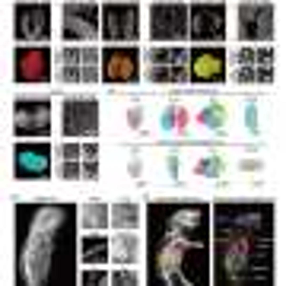
Traditional pathology relies on thin, two‑dimensional slices, which obscure the spatial relationships that drive organ function and disease. Recent advances in tissue‑clearing, particularly the CUBIC protocol, have enabled researchers to render opaque samples transparent while preserving cellular integrity. Coupled with light‑sheet fluorescence microscopy, these methods generate high‑resolution, volumetric datasets that capture the exact location of each nucleus, laying the groundwork for true whole‑organ and whole‑body imaging.
The University of Tokyo team leveraged this technology to construct the CUBIC Organ/Body Atlas, a cell‑by‑cell map of a neonatal mouse. By extracting positional data for millions of cells, they created a reference framework that can be superimposed with experimental datasets, allowing direct comparison of cellular distributions under different genetic or pharmacological conditions. This quantitative capability is a game‑changer for developmental biology, where researchers can now track organogenesis at single‑cell resolution, and for drug discovery, where off‑target effects can be assessed across the entire organism rather than isolated tissues.
Looking ahead, integrating these 3D atlases with spatial transcriptomics and other omics layers will deliver a unified view of morphology and molecular activity. Such multimodal maps could underpin next‑generation diagnostic platforms that surpass the limitations of conventional histology, offering clinicians a holistic picture of disease spread. As the technology matures and adapts to larger, human tissues, it is poised to create new markets in precision pathology, biotech research services, and AI‑driven image analysis, reshaping how the biomedical industry approaches disease characterization and therapeutic evaluation.
From slices to whole bodies: How 3D cell atlases could reshape pathology research
Comments
Want to join the conversation?
Loading comments...