New 3D Microscope Technology Captures High-Resolution Tissue Images at a Fraction of the Cost
Why It Matters
HySIL breaks the cost‑performance trade‑off in 3D microscopy, making high‑resolution tissue imaging affordable for research labs and clinical settings, thereby accelerating AI‑driven pathology and neuroscience breakthroughs.
Key Takeaways
- •HySIL merges solid lens with immersion liquid for high resolution
- •SCOPE retrofit adds HySIL to existing light‑sheet microscopes
- •Costs drop to a fraction of traditional oil‑immersion systems
- •Enables centimeter‑scale 3D imaging for labs, clinics, and AI training
Pulse Analysis
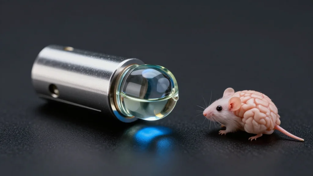
The hybrid solid‑liquid optics (HySIL) platform reimagines how light is captured in microscopy. By pairing a simple curved glass lens with a precisely matched immersion liquid, the system creates a continuous optical path that rivals expensive oil‑immersion objectives while using inexpensive air lenses. This clever redesign eliminates the need for specialized hardware, allowing the modular SCOPE attachment to slot onto standard light‑sheet microscopes and even other modalities such as confocal or two‑photon systems. The result is sub‑micron resolution across tissue depths of several centimeters, a scale previously limited to costly, custom‑built instruments.
Beyond the engineering feat, HySIL’s affordability and ease of integration open new avenues for large‑scale biological imaging. Researchers can now generate dense 3D datasets from whole organs, cancer biopsies, and organoid models without prohibitive expense. These rich image volumes are ideal training material for deep‑learning algorithms that aim to automate disease detection, grading, and prognostication. By democratizing access to high‑quality volumetric data, the technology accelerates the feedback loop between experimental biology and AI‑driven diagnostics, potentially shortening the path from discovery to clinical application.
Commercially, the technology is already moving from the lab to the market. MBF Bioscience, a long‑time partner, has incorporated HySIL into its SLICE light‑sheet system, offering a turnkey solution for teaching labs, research institutions, and low‑resource clinics. Patent filings by Columbia University protect the core optics and modular designs, positioning the team for licensing and further spin‑outs. As the demand for scalable, high‑resolution imaging grows across neuroscience, developmental biology, and pathology, HySIL could become a standard component in the next generation of microscopy platforms.
New 3D microscope technology captures high-resolution tissue images at a fraction of the cost
Comments
Want to join the conversation?
Loading comments...