Structural Covariance, Regional Topology, and Volumetric Aspects of Amygdala Subnuclei in Posttraumatic Stress Disorder Using Ultra-High Field Imaging
•December 29, 2025
0
Why It Matters
The findings pinpoint amygdala subnucleus alterations as potential neurobiological markers of PTSD risk and trauma resilience, informing precision diagnostics and targeted interventions.
Key Takeaways
- •Lateral nucleus volume distinguishes PTSD from non‑trauma controls
- •Right paralaminar nucleus shows higher connectivity in trauma‑exposed controls
- •Structural covariance patterns differ across PTSD, trauma‑exposed, and healthy groups
- •No whole amygdala volume differences detected
- •7‑Tesla MRI enables subnucleus‑level analysis
Pulse Analysis
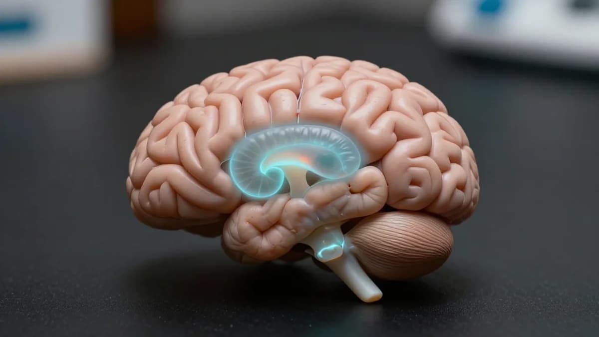
The amygdala has long been treated as a monolithic structure in psychiatric imaging, yet its nine subnuclei serve distinct functional roles in fear, memory, and reward processing. Conventional 3‑Tesla scanners lack the spatial resolution to reliably separate these tiny regions, leading to mixed volumetric findings in PTSD research. By leveraging 7‑Tesla ultra‑high‑field MRI, the study achieved sub‑millimetre resolution, allowing precise segmentation of each subnucleus and revealing nuanced volume changes that were previously obscured. This methodological advance underscores the importance of high‑field imaging for dissecting complex neurocircuitry in mental‑health disorders.
Beyond size, the investigation applied graph‑theory to map how each subnucleus covaries structurally with the rest of the brain. Trauma‑exposed individuals without PTSD exhibited a heightened nodal degree in the right paralaminar nucleus, suggesting a compensatory hub that may support adaptive processing of salient stimuli. In contrast, PTSD participants did not show such topological enhancements, implying a disruption of network integration that could underlie hyper‑reactivity to threat cues. These topology insights complement traditional functional connectivity studies, offering a structural counterpart to the dynamic dysregulation observed in PTSD.
The most striking outcomes emerged from the structural‑covariance analyses, where specific subnucleus‑region pairings differed across groups. Non‑trauma controls displayed stronger covariance between the left central and cortical nuclei with the hippocampus, aligning with intact memory consolidation pathways. Conversely, trauma‑exposed controls showed reduced covariance between several nuclei and medial temporal structures, hinting at a re‑wiring that may buffer against symptom development. Such subregional signatures provide a fertile ground for biomarker development, guiding future longitudinal studies and personalized therapeutic strategies that target the most vulnerable amygdala circuits.
Structural covariance, regional topology, and volumetric aspects of amygdala subnuclei in posttraumatic stress disorder using ultra-high field imaging
0
Comments
Want to join the conversation?
Loading comments...