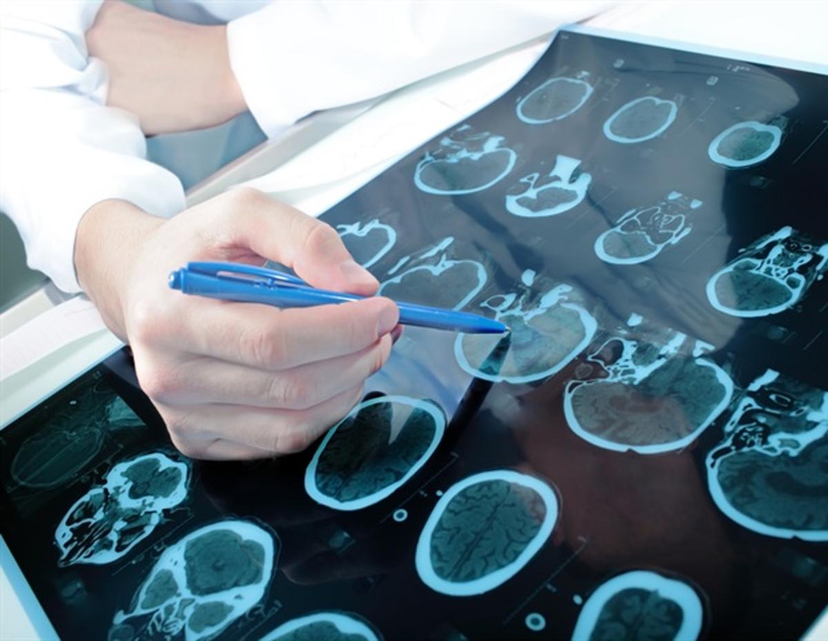
Deep Learning Model Predicts Vascular Cognitive Impairment From Brain Scans
Why It Matters
The method gives clinicians an objective, imaging‑based tool to diagnose and profile vascular cognitive deficits without lengthy neuropsych testing, accelerating precision‑medicine interventions. Its high accuracy across scanners suggests scalability for broader clinical adoption.
Key Takeaways
- •DenseNet achieved 90.2% internal accuracy, 92.6% on external data
- •Model predicts cognitive scores, correlating with MoCA, MMSE, Trail Making
- •Salient maps highlight 11 white‑matter tracts linked to vascular injury
- •SSIM clustering stratifies patients by domain‑specific cognitive risk
- •Framework uses only standard DTI, eliminating need for neuropsych testing
Pulse Analysis
Vascular contributions to cognitive decline are increasingly recognized as a major cause of dementia, yet distinguishing subcortical vascular cognitive impairment (SVCI) from other small‑vessel pathologies remains difficult. Conventional MRI reveals white‑matter hyperintensities, but these lesions are also common in healthy aging, limiting diagnostic specificity. Diffusion tensor imaging (DTI) captures microstructural changes through fractional anisotropy and mean diffusivity, offering a more sensitive window into white‑matter integrity. However, interpreting high‑dimensional DTI data requires expertise and time, creating a clear opportunity for automated, data‑driven solutions.
The research team built a densely connected convolutional network (DenseNet) that ingests raw FA and MD volumes and learns hierarchical features without manual engineering. After training on 305 internal subjects, unsupervised domain adaptation allowed the model to maintain performance on an independent cohort from different scanners, achieving 90.2% internal accuracy and 92.6% external accuracy with AUCs above 0.94. Guided backpropagation produced voxel‑wise saliency maps that consistently highlighted eleven white‑matter tracts—such as the corona radiata and corpus callosum—known to underlie attention and memory, confirming biological plausibility.
Beyond binary classification, the authors introduced a novel SSIM‑based clustering that matches each patient’s saliency pattern to domain‑specific relevance maps, effectively generating a personalized cognitive‑risk signature from a single DTI scan. This eliminates the need for lengthy neuropsychological batteries, making the tool attractive for busy neurology clinics and resource‑limited settings. Ongoing longitudinal follow‑up of the VIVA cohort will test the model’s ability to predict future decline, while future work aims to fuse multimodal imaging and blood biomarkers, moving vascular cognitive impairment closer to true precision medicine.
Deep learning model predicts vascular cognitive impairment from brain scans
Comments
Want to join the conversation?
Loading comments...