Diffusion MRI as A Biomarker for Monitoring Recovery After Surgical Repair of Traumatic Peripheral Nerve Injuries: A Longitudinal Case Series
Why It Matters
Accurate, early detection of nerve repair success can reduce repeat surgeries and improve functional outcomes, addressing a major gap in peripheral nerve trauma care.
Key Takeaways
- •Fractional anisotropy (FA) values tracked nerve recovery over 12 months
- •FA trends aligned with Medical Research Council sensory and motor scores
- •Gompertz model automated analysis, reducing user‑dependent interpretation variability
- •Study focused on severe ulnar and median nerve transections
- •Noninvasive DTI offers potential early detection of repair failure
Pulse Analysis
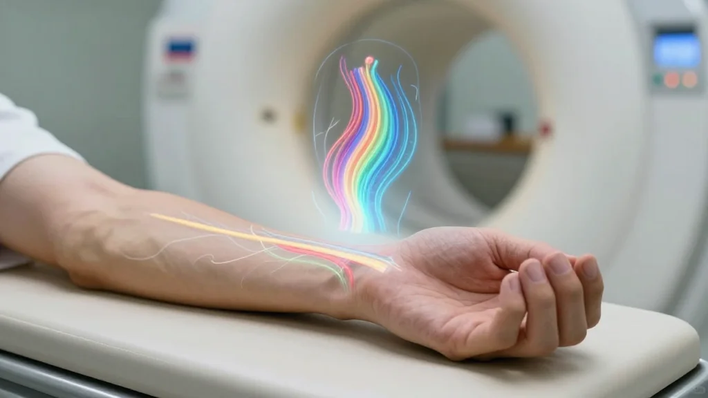
Peripheral nerve injuries, especially transections of the ulnar and median nerves, present a daunting clinical challenge because delayed or failed regeneration often leads to permanent disability. Traditional assessment relies on serial physical examinations and electromyography, which can miss early signs of inadequate repair. In this context, diffusion tensor imaging (DTI) emerges as a promising imaging biomarker, capturing microstructural water diffusion along axonal pathways. By quantifying fractional anisotropy, clinicians gain a window into the integrity of regenerating fibers, offering a more objective and timely metric than subjective sensory or motor grading alone.
The study leveraged a longitudinal design, acquiring DTI scans at multiple intervals over a year after surgical repair. Researchers applied a Gompertz function—a sigmoidal growth model—to the FA data, automatically characterizing the nonlinear recovery curve. This approach not only aligned closely with the Medical Research Council’s sensory and motor scales but also eliminated much of the inter‑observer variability that hampers conventional image interpretation. The automated pipeline demonstrated that FA increases reliably signal functional improvement, while stagnation or decline may flag impending repair failure, prompting earlier clinical decision‑making.
If integrated into routine postoperative protocols, FA‑based DTI could transform peripheral nerve surgery by enabling proactive management. Surgeons could identify suboptimal regeneration weeks rather than months after the procedure, reducing the need for costly revision surgeries and associated morbidity. Moreover, the technology opens avenues for pharmaceutical trials targeting nerve regeneration, providing a quantifiable endpoint. As imaging hardware becomes more accessible and analysis algorithms mature, the market for neuro‑imaging biomarkers is poised for rapid growth, positioning DTI as a cornerstone of precision neuro‑rehabilitation.
Diffusion MRI as A Biomarker for Monitoring Recovery After Surgical Repair of Traumatic Peripheral Nerve Injuries: A Longitudinal Case Series
Comments
Want to join the conversation?
Loading comments...