Researchers Use Multi-Modality Imaging to Learn More About MINOCA
Why It Matters
The findings demonstrate that MINOCA is not a benign diagnosis and that advanced imaging can guide targeted therapy, potentially reducing the substantial long‑term cardiovascular risk.
Key Takeaways
- •Multi-modality imaging identified causes in 79% of MINOCA patients
- •True myocardial infarction present in 59% of studied cohort
- •Mimicking conditions accounted for 20% of cases, detected via MRI
- •No significant sex differences in underlying mechanisms found
- •One‑year major adverse event rate for MINOCA is 10%
Pulse Analysis
MINOCA, or myocardial infarction with nonobstructive coronary arteries, has long perplexed cardiologists because patients present with classic heart‑attack symptoms yet show no significant blockages on angiography. Historically, the condition was more frequently reported in women, leading to the belief that sex‑specific mechanisms drive its pathology. However, the prevalence of MINOCA in the broader population is rising as clinicians adopt more sensitive diagnostic tools, making it a critical focus for cardiovascular research and practice.
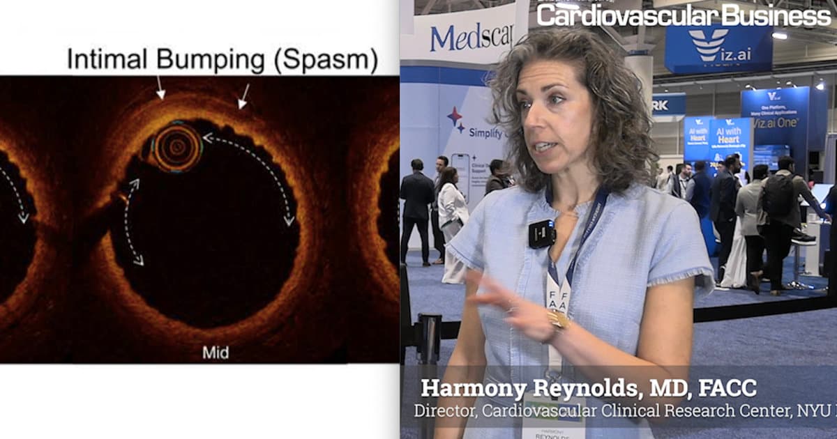
The recent NYU Langone study leveraged high‑resolution optical coherence tomography (OCT) alongside cardiac magnetic resonance imaging (MRI) to interrogate both the coronary vessel wall and myocardial tissue. By pairing these modalities, investigators pinpointed an underlying cause in nearly eight out of ten patients, revealing that true myocardial infarction accounts for 59% of cases while 20% are attributable to conditions that mimic heart attacks, such as myocarditis. Importantly, the data showed no statistically significant differences between men and women in the distribution of these mechanisms, suggesting that treatment strategies should be guided by imaging findings rather than gender alone.
Clinically, the research carries weighty implications. MINOCA patients face a 10% one‑year and 24% four‑year risk of major adverse cardiovascular events, contradicting the outdated practice of discharging them without therapy. The study advocates for routine use of OCT and MRI in suspected MINOCA cases to uncover treatable etiologies—be it plaque rupture, coronary spasm, or microvascular dysfunction—and to tailor secondary‑prevention regimens accordingly. As guidelines evolve, incorporating multi‑modality imaging could become the standard of care, improving outcomes for a condition that has historically been under‑diagnosed and under‑treated.
Researchers use multi-modality imaging to learn more about MINOCA
Comments
Want to join the conversation?
Loading comments...