Using Tiny Ripples at Skin Level to Monitor for Possible Health Problems Below
•March 5, 2026
0
Why It Matters
The approach transforms ubiquitous mobile cameras into diagnostic tools, potentially democratizing early detection of conditions like tumors or liver disease and reducing reliance on costly imaging equipment.
Key Takeaways
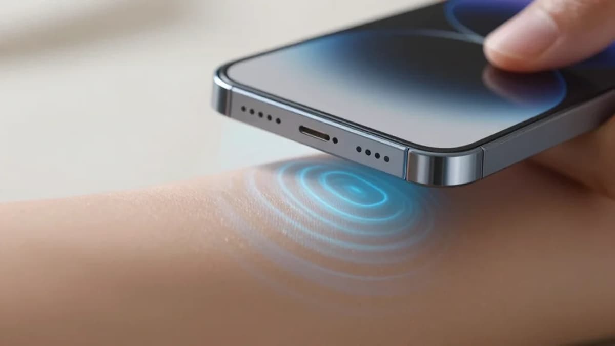
- •Visual surface wave elastography uses smartphone video to assess tissue.
- •Detects subpixel motion as small as 1/500 pixel.
- •Measures stiffness and thickness, biomarkers for tumors, liver disease.
- •Validated against rheometer and simulated leg models.
- •Enables low-cost, at‑home health monitoring without contact.
Pulse Analysis
The emergence of visual surface wave elastography marks a convergence of computer vision and biomedical diagnostics, leveraging everyday devices to probe beneath the skin. By detecting minute surface ripples generated by simple stimuli—such as a massage gun or ambient sound—the algorithm translates visual data into quantitative measures of tissue stiffness and thickness. This non‑contact method sidesteps the need for traditional ultrasound transducers or MRI scanners, offering a scalable solution for continuous health tracking.
From a technical perspective, the breakthrough hinges on phase‑based motion processing that resolves subpixel displacements down to 0.002 pixels. Coupled with spectral decomposition, researchers construct a dispersion relation that maps wave frequency and wavenumber to underlying material properties. Validation against high‑precision rheometers and anatomically accurate simulations demonstrates that the technique can reliably differentiate between healthy and pathological tissue states, even on irregular body geometries. Such precision opens pathways for early biomarker detection, including tumor stiffening and liver fibrosis progression.
Clinically, the implications are profound. A smartphone‑based elastography platform could empower patients to perform routine self‑assessments, generating longitudinal data that alerts clinicians to subtle changes before symptoms arise. This democratization of diagnostic imaging promises to alleviate pressure on healthcare systems, especially in underserved regions where access to advanced imaging is limited. As the technology matures, integration with telemedicine workflows and AI‑driven interpretation could further streamline early intervention strategies, reshaping preventive medicine.
Using tiny ripples at skin level to monitor for possible health problems below
0
Comments
Want to join the conversation?
Loading comments...