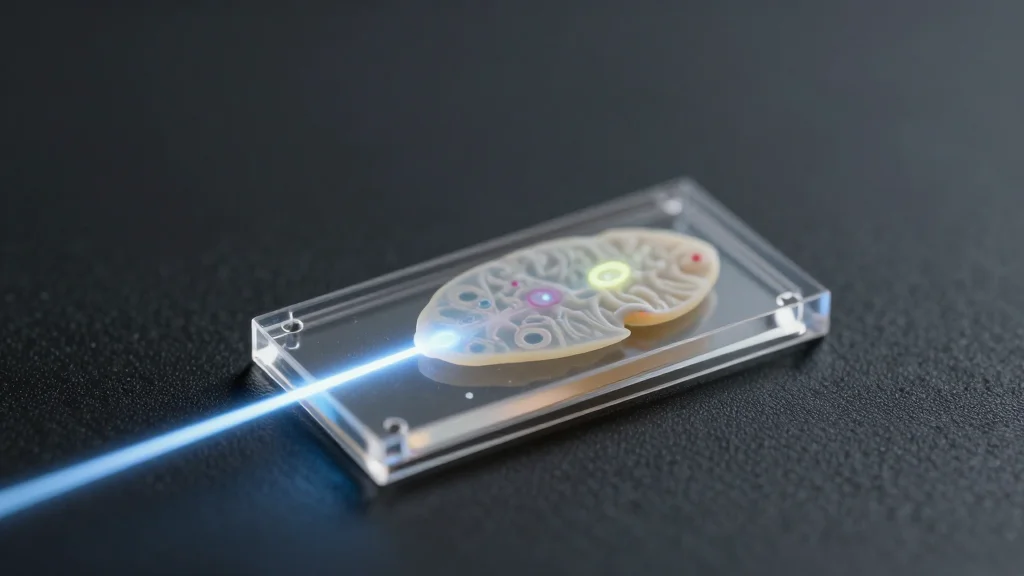
Pencil Beam Laser Could Help Researchers Design Brain-Targeted Therapies
Why It Matters
The ability to image drug penetration across the blood‑brain barrier at cellular resolution and unprecedented speed could dramatically shorten neuro‑drug development cycles and improve predictive accuracy over animal models.
Key Takeaways
- •MIT team discovered self‑organizing laser pencil beam for bioimaging
- •Technique captures 3D blood‑brain barrier images 25× faster than standard
- •No fluorescent tags needed, enabling real‑time drug uptake tracking
- •High‑resolution, deep‑focus beam overcomes traditional imaging trade‑offs
- •Could accelerate neuro‑drug development and broader tissue‑engineered model studies
Pulse Analysis
The breakthrough stems from a counter‑intuitive optical physics insight: under specific conditions, scattered laser photons can spontaneously self‑align into a narrow, stable pencil beam rather than devolving into chaos. By harnessing this self‑localization, MIT’s EECS lab sidestepped the usual trade‑off between power and image fidelity, delivering a beam that maintains high resolution across a deep focal range. This advance not only pushes the limits of multiphoton microscopy but also signals a paradigm shift for researchers who rely on precise, high‑speed imaging in complex biological systems.
Applied to the human blood‑brain barrier, the new technique generated volumetric, cellular‑level images up to 25 times faster than conventional two‑photon methods. Crucially, it does so without requiring fluorescent labeling, allowing scientists to watch drug molecules enter endothelial cells in real time. This capability addresses a long‑standing bottleneck in neuropharmaceutical research, where animal models often misrepresent human drug permeability. By providing a rapid, label‑free readout of drug uptake, the method promises to streamline screening pipelines for Alzheimer’s, ALS, and other neurodegenerative conditions, reducing both cost and time to market.
Beyond the immediate biomedical application, the self‑organized pencil beam could become a versatile tool across tissue‑engineered platforms, from organ‑on‑a‑chip systems to in‑situ neuronal imaging. Industry stakeholders are already eyeing commercialization pathways, recognizing the competitive edge of faster, higher‑resolution imaging for drug discovery and diagnostics. Future work will deepen the physical understanding of the beam’s self‑organization and expand its use cases, potentially reshaping how researchers visualize dynamic molecular processes in living tissues. The convergence of optical physics and translational medicine embodied in this work underscores the growing importance of interdisciplinary innovation in the biotech sector.
Pencil Beam Laser Could Help Researchers Design Brain-Targeted Therapies
Comments
Want to join the conversation?
Loading comments...