New Cytometer Measures Cell Stiffness to Improve Disease Diagnosis
Why It Matters
High‑throughput mechanical phenotyping offers a rapid, label‑free biomarker that can accelerate disease diagnosis and prognosis. It also reduces reliance on costly, low‑throughput AFM equipment, paving the way for clinical adoption.
Key Takeaways
- •Measures cell stiffness using time‑of‑flight in microfluidic channels
- •Processes 60–100 cells per second, orders of magnitude faster than AFM
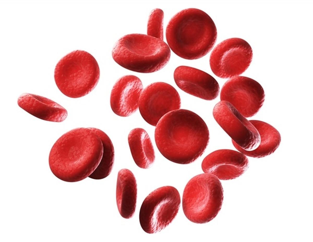
- •Distinguishes soft cancer cells from stiff malaria‑affected red blood cells
- •Collaboration combines Brown’s synthetic particles with NIST’s cytometer design
- •Aims to enable high‑throughput diagnostic testing of patient samples
Pulse Analysis
The mechanical properties of cells have long been recognized as a window into disease biology, yet their measurement has remained a niche technique. Traditional atomic force microscopy (AFM) provides nanometer‑scale elasticity data but requires cells to be immobilized and examined one at a time, limiting throughput to roughly one cell every 30 seconds. This bottleneck prevents large‑scale studies and hampers translation into clinical workflows, where rapid, label‑free biomarkers are essential for timely diagnosis of cancers, infectious diseases, and hematologic disorders.
The Brown University–NIST team sidestepped these constraints by repurposing a conventional flow cytometer into a mechanophenotyping platform. By recording the time‑of‑flight of each cell as it traverses a microfluidic channel, the device infers stiffness: softer cells migrate toward the fast‑moving core, while stiffer cells linger near slower peripheral streams. Leveraging existing fluorescence detectors for size measurement, the system simultaneously captures physical and biochemical signatures. In proof‑of‑concept tests it resolved particles of differing elastic moduli at rates of 60–100 cells per second, with potential scaling to thousands per second.
Because mechanical phenotype can differentiate malignant from benign cells and flag infections such as malaria or sickle‑cell disease, the cytometer promises a new layer of diagnostic information that complements genetic and protein‑based assays. Its high throughput and compatibility with standard flow‑cytometry hardware could accelerate adoption in clinical labs, reducing reliance on costly AFM setups. Ongoing collaborations with Brown’s medical partners aim to validate the technology on patient blood and tissue samples, a step that could open commercial pathways for point‑of‑care devices and broaden the market for microfluidic diagnostics.
New cytometer measures cell stiffness to improve disease diagnosis
Comments
Want to join the conversation?
Loading comments...