SNMMI Unveils 2026 Image of the Year
Why It Matters
Direct clot visualization could replace several conventional tests, reducing time, cost, and radiation exposure while improving treatment decisions for vascular disease.
Key Takeaways
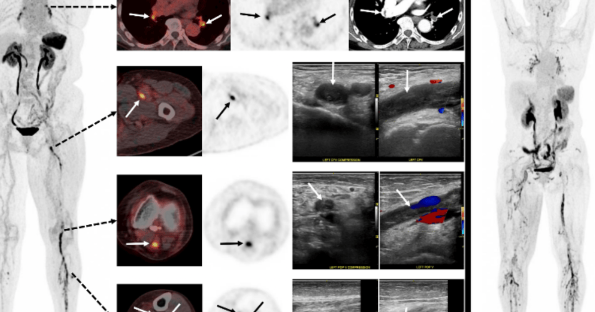
- •¹⁸F‑GP1 PET/CT directly visualizes thrombi, surpassing ultrasound limitations
- •Whole-body imaging detects DVT and concurrent pulmonary embolism in one scan
- •Tracer showed high diagnostic accuracy comparable to standard ultrasound
- •No adverse events reported, indicating strong patient tolerability
- •Potential platform for imaging strokes and broader cardiovascular disease
Pulse Analysis
The selection of a whole‑body PET image as SNMMI’s 2026 Image of the Year underscores a turning point in vascular diagnostics. Traditional modalities such as duplex ultrasound and contrast‑enhanced CT rely on indirect signs—vein compressibility or filling defects—to infer clot presence. By contrast, molecular imaging with targeted radiotracers can illuminate the clot itself, offering clinicians a direct view of platelet activation. This shift aligns with a broader industry trend toward precision imaging, where functional information complements anatomy to improve decision‑making. Such clarity can also guide anticoagulant therapy choices.
The investigational tracer ¹⁸F‑GP1 targets activated platelets, enabling PET/CT scans to pinpoint deep‑vein thrombosis and simultaneous pulmonary embolism. In a multicenter study, its sensitivity and specificity matched those of duplex ultrasound for thigh and calf clot detection, while also revealing emboli that would otherwise require a separate CT pulmonary angiogram. Patients tolerated the agent without any reported adverse events, a critical factor for widespread clinical adoption. By consolidating multiple examinations into a single whole‑body scan, the technology promises faster diagnosis and reduced radiation exposure.
Beyond venous disease, experts envision ¹⁸F‑GP1 as a platform for detecting arterial thrombi, stroke‑related clots, and even atherosclerotic plaque activity. If integrated into routine workflows, the tracer could eliminate the need for separate ultrasound, CT, or MRI studies, streamlining patient pathways and cutting hospital costs. Pharmaceutical firms are already exploring similar platelet‑targeted agents, suggesting a burgeoning market for thrombus‑specific imaging. As reimbursement frameworks adapt to value‑based care, the ability to provide comprehensive, single‑session assessments may accelerate adoption across cardiology, oncology, and emergency medicine.
SNMMI unveils 2026 Image of the Year
Comments
Want to join the conversation?
Loading comments...