3D Microscopy Reveals How a Tick-Borne Virus Reshapes Human Cells to Replicate
Why It Matters
Understanding TBEV’s cellular remodeling uncovers novel antiviral targets, accelerating drug development against a growing public‑health threat. This knowledge could improve treatment strategies for tick‑borne encephalitis worldwide.
Key Takeaways
- •Tick-borne encephalitis virus reorganizes host cell membranes.
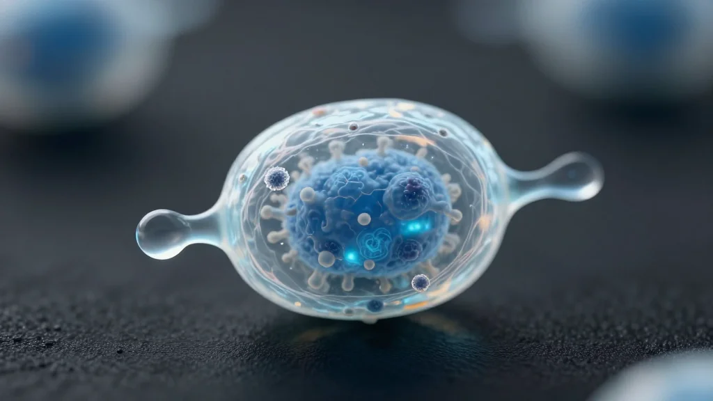
- •3D microscopy visualized viral replication complexes.
- •Virus creates specialized “factory” compartments for assembly.
- •Findings reveal potential antiviral targets in replication pathway.
- •Study published in Nature Communications, Umeå University.
Pulse Analysis
Tick-borne encephalitis (TBE) has emerged as a significant vector‑borne disease across Europe and Asia, with incidence rates climbing due to expanding tick habitats and climate change. Despite widespread vaccination programs, therapeutic options remain limited, leaving clinicians reliant on supportive care. The new study arrives at a critical juncture, offering a molecular‑level view of how the TBE virus commandeers host cell architecture, a gap that has hampered rational drug design for years.
The research team employed cutting‑edge three‑dimensional structured illumination microscopy, achieving nanometer‑scale resolution of infected human fibroblasts. Images captured the virus‑induced reshaping of endoplasmic reticulum membranes into tightly curved vesicles and tubular networks that serve as hubs for RNA replication and virion assembly. By correlating these structures with viral protein markers, the scientists demonstrated that TBEV orchestrates a coordinated remodeling program, effectively converting ordinary cytoplasm into a dedicated viral factory. Such visual evidence substantiates prior biochemical models and provides a concrete framework for targeting the replication complex.
These insights have immediate translational relevance. The identified membrane‑bound compartments expose virus‑specific enzymes and scaffolding proteins that are absent in uninfected cells, presenting attractive candidates for small‑molecule inhibitors. Moreover, the methodological blueprint can be extended to other flaviviruses, such as West Nile and Zika, accelerating cross‑species antiviral research. As the scientific community seeks to curb the public‑health burden of tick‑borne illnesses, this work equips drug developers with actionable structural targets and underscores the value of high‑resolution imaging in infectious‑disease biology.
3D microscopy reveals how a tick-borne virus reshapes human cells to replicate
Comments
Want to join the conversation?
Loading comments...