Astrocytes Connect Specific Brain Regions Through Plastic Networks
Why It Matters
The work shows astrocyte gap‑junction networks are organized, region‑specific, and adaptable, positioning them as active participants in neural circuitry and potential therapeutic targets for neurodegenerative and neurodevelopmental disorders.
Key Takeaways
- •Cx43‑TurboID viral tracer maps astrocyte gap‑junction networks in mice
- •Motor, prefrontal and hypothalamic astrocytes form distinct, conserved subnetworks
- •Dual Cx43/Cx30 knockout collapses network spread, confirming gap‑junction dependence
- •Whisker trimming reduces astrocyte connectivity, revealing network plasticity
Pulse Analysis
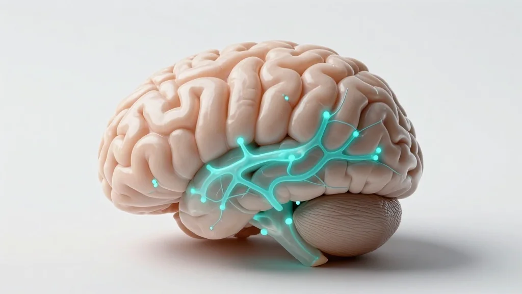
Astrocytes have long been viewed as supportive glial cells, yet their ability to share metabolites and signals through gap‑junction channels is now recognized as a cornerstone of central nervous system homeostasis. Gap‑junctions, primarily composed of connexin‑43 (Cx43) and connexin‑30 (Cx30), create a syncytial web that can redistribute ions, neurotransmitters, and energy substrates across vast brain territories. Understanding the spatial layout of this web has been hampered by techniques that either disrupt native connectivity or capture only local interactions, leaving a critical gap in our knowledge of how astrocytes contribute to brain‑wide signaling.
The new study overcomes these limitations by engineering an adeno‑associated virus that expresses a Cx43‑TurboID fusion protein under a glial‑specific promoter. When incorporated into gap‑junction hemichannels, TurboID biotinylates molecules traversing the junction, allowing researchers to visualize infected astrocytes (HA‑positive) and their connected partners (streptavidin‑positive) without background noise. In vivo light‑sheet imaging of mice injected in the motor cortex, prefrontal cortex, or hypothalamus revealed highly organized, region‑specific subnetworks that are remarkably consistent across individuals. Importantly, mice lacking both Cx43 and Cx30 showed a dramatic collapse of these networks, confirming that the observed connectivity relies exclusively on astrocytic gap‑junctions rather than alternative pathways such as vascular routes.
Beyond mapping, the work demonstrates functional plasticity: trimming whiskers—a classic model of sensory deprivation—significantly reduced the proportion of biotin‑labeled astrocytes in the corresponding barrel cortex. This suggests that astrocyte networks can remodel in response to experience, mirroring neuronal plasticity. The findings open new avenues for exploring how astrocytic connectivity influences learning, memory, and disease progression, and they provide a versatile tool for probing glial contributions to brain function in health and pathology.
Astrocytes connect specific brain regions through plastic networks
Comments
Want to join the conversation?
Loading comments...