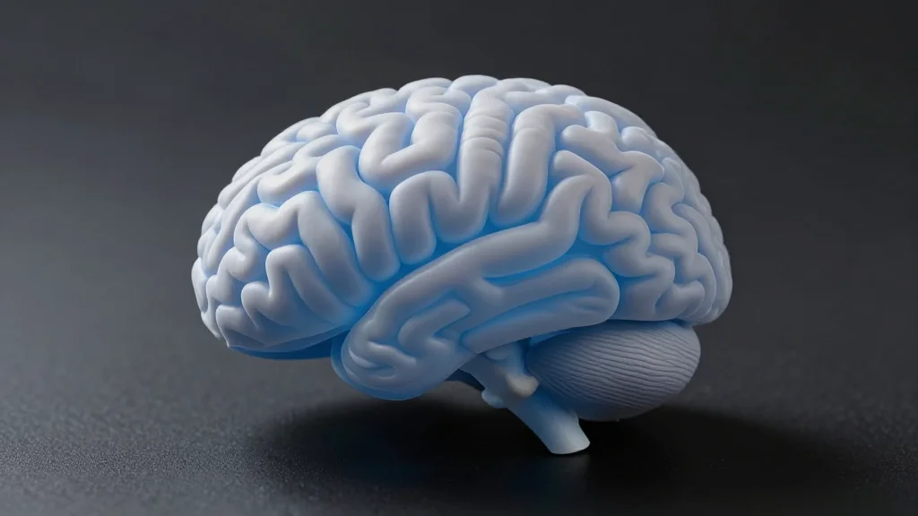
Brain Scans of 800 Incarcerated Men Link Psychopathy to an Expanded Cortical Surface Area
Why It Matters
The findings provide a more precise neurobiological marker for psychopathic traits, which could improve risk assessment and inform targeted interventions in forensic and clinical settings.
Key Takeaways
- •804 incarcerated men with high psychopathy show larger cortical surface area.
- •Expanded surface area concentrated in superior temporal, auditory, and paralimbic regions.
- •Compressed cortical thickness gradients indicate reduced segregation of sensory and associative networks.
- •Self-reported empathy scores showed no significant link to brain structure.
Pulse Analysis
Recent advances in neuroimaging have begun to untangle the complex biology underlying psychopathy, a condition marked by shallow affect, manipulative behavior, and impulsivity. Traditional studies focused on overall gray‑matter volume, often reporting reductions in brain size among psychopathic individuals. However, cortical surface area and thickness develop through distinct genetic and cellular pathways, meaning that lumping them together can mask nuanced patterns. By isolating surface area, researchers can capture developmental processes such as neuronal migration and cortical folding that may be uniquely altered in antisocial phenotypes.
The new investigation leveraged a mobile MRI unit to scan 804 adult men across multiple correctional facilities, creating one of the largest neuroanatomical datasets for this population. Using the Psychopathy Checklist‑Revised, the team identified a clear association between high psychopathy scores and an expanded cortical surface, particularly in the superior temporal cortex, auditory regions, and paralimbic structures that bridge emotion and cognition. Simultaneously, the gradient of cortical thickness—a map that normally stretches from primary sensory zones to higher‑order association areas—was noticeably compressed, suggesting a blurring of functional specialization. Notably, these brain metrics did not correlate with self‑reported empathy, highlighting a disconnect between subjective assessments and underlying neural architecture.
These insights have practical ramifications for both forensic psychology and public health. A reliable structural marker could refine risk‑assessment tools, aiding parole boards and clinicians in identifying individuals who may benefit from early, intensive interventions. The study also flags methodological gaps: reliance on self‑report empathy scales and an exclusively male, incarcerated sample limit generalizability. Future work should incorporate performance‑based empathy tasks, diversify participant demographics, and explore the cellular mechanisms driving cortical expansion. Such research could eventually inform preventive strategies aimed at fostering empathy and reducing severe antisocial behavior before it manifests.
Brain scans of 800 incarcerated men link psychopathy to an expanded cortical surface area
Comments
Want to join the conversation?
Loading comments...