Electrospinning of Hydroxypropyl Chitosan Nanofibers for Bone Regeneration Application
Why It Matters
The finding offers a scalable, biocompatible scaffold that could accelerate bone healing, addressing a critical need in orthopedic implants and tissue engineering.
Key Takeaways
- •50/50 HPCH‑PVA fibers boost osteoblast differentiation
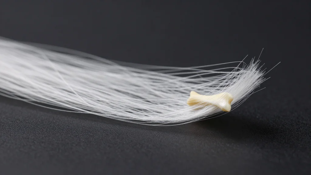
- •SEM and FT‑IR confirm uniform nanofiber morphology
- •Alizarin Red staining shows increased mineral deposition
- •Scaffold combines biodegradability with mechanical stability
Pulse Analysis
Electrospinning has emerged as a versatile technique for fabricating nanofibrous scaffolds that mimic the extracellular matrix of bone tissue. Traditional materials such as pure collagen or synthetic polymers often struggle to balance mechanical strength with bioactivity, leading researchers to explore hybrid polymers. Hydroxypropyl chitosan (HPCH), a water‑soluble derivative of chitosan, offers inherent osteoconductivity and biodegradability, while poly(vinyl alcohol) (PVA) contributes tensile strength and processability. By adjusting polymer ratios, scientists can fine‑tune fiber diameter, porosity, and degradation rates, which are critical parameters for supporting cell attachment and nutrient diffusion.
In the recent study, a 50 % HPCH‑50 % PVA formulation produced nanofiber mats with consistent morphology, as verified by scanning electron microscopy and Fourier‑transform infrared spectroscopy. Mechanical testing indicated sufficient elasticity to withstand handling, yet the scaffold remained soft enough to permit cellular infiltration. When cultured with MC3T3 pre‑osteoblasts, the hybrid fibers triggered a marked increase in alkaline phosphatase activity and Alizarin Red staining, signaling enhanced mineralization. Gene expression analysis further revealed up‑regulation of key osteogenic markers such as Runx2 and OCN, confirming that the scaffold actively promotes differentiation rather than serving merely as a passive substrate.
The implications for the orthopedic and regenerative‑medicine markets are significant. A cost‑effective, easily scalable HPCH‑PVA scaffold could reduce reliance on autografts and expensive synthetic implants, shortening recovery times and lowering complication rates. Moreover, the material’s tunable degradation profile aligns with the timeline of natural bone healing, potentially eliminating the need for secondary surgeries to remove hardware. Future work will likely focus on in‑vivo validation, integration with growth‑factor delivery systems, and regulatory pathways, positioning this technology for rapid translation into clinical practice.
Electrospinning of hydroxypropyl chitosan nanofibers for bone regeneration application
Comments
Want to join the conversation?
Loading comments...