Fetal Brain Scans Can Predict a Toddler’s Vocabulary Size Years Before They Learn to Speak
Why It Matters
Prenatal brain metrics could become early indicators for language outcomes, informing interventions before speech delays manifest. This bridges a gap between fetal neurodevelopment research and practical pediatric screening.
Key Takeaways
- •Prenatal superior temporal gyrus volume predicts toddler vocabulary size
- •Prediction holds for both left and right brain hemispheres
- •Inferior frontal gyrus size shows no link to early word count
- •Study limited to 41 scans, 24 children in final analysis
- •Findings based on predominantly white, high‑income families
Pulse Analysis
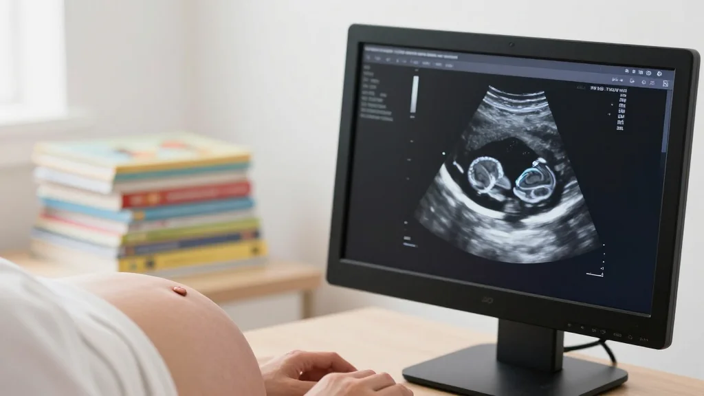
Understanding language acquisition has long focused on post‑natal brain growth, yet the new findings from the Max Planck Institute suggest that the groundwork is laid well before birth. The superior temporal gyrus, a region critical for processing auditory signals, begins forming around the 24‑week mark and reaches a measurable size by the third trimester. By capturing high‑resolution fetal MRI scans during weeks 30‑33, researchers can now quantify subtle variations that later correlate with expressive vocabulary, offering a rare glimpse into the prenatal origins of linguistic ability.
The study followed 41 fetuses through the Cambridge Human Imaging and Longitudinal Development (CHILD) project, ultimately analyzing language outcomes for 24 toddlers aged 24‑36 months. Children with larger bilateral superior temporal gyri produced significantly more words, a relationship that emerged only after the second year of life, suggesting that early neural architecture sets a ceiling for later lexical growth. In contrast, the inferior frontal gyrus—associated with grammar and higher‑order language processing—did not predict early word counts, underscoring the distinct developmental timelines of language‑related brain regions.
While the sample size and demographic homogeneity limit generalizability, the research opens avenues for early‑risk screening and targeted interventions. If prenatal imaging can reliably flag children at risk for delayed language, clinicians could deploy enrichment programs well before traditional milestones are missed. Future work must expand to diverse populations and incorporate receptive vocabulary and other linguistic domains, but the current evidence positions fetal brain metrics as a promising tool in the preventive pediatric toolkit.
Fetal brain scans can predict a toddler’s vocabulary size years before they learn to speak
Comments
Want to join the conversation?
Loading comments...