High-Resolution Imaging Shines Light on Nanoscale Nuclear Organization
Why It Matters
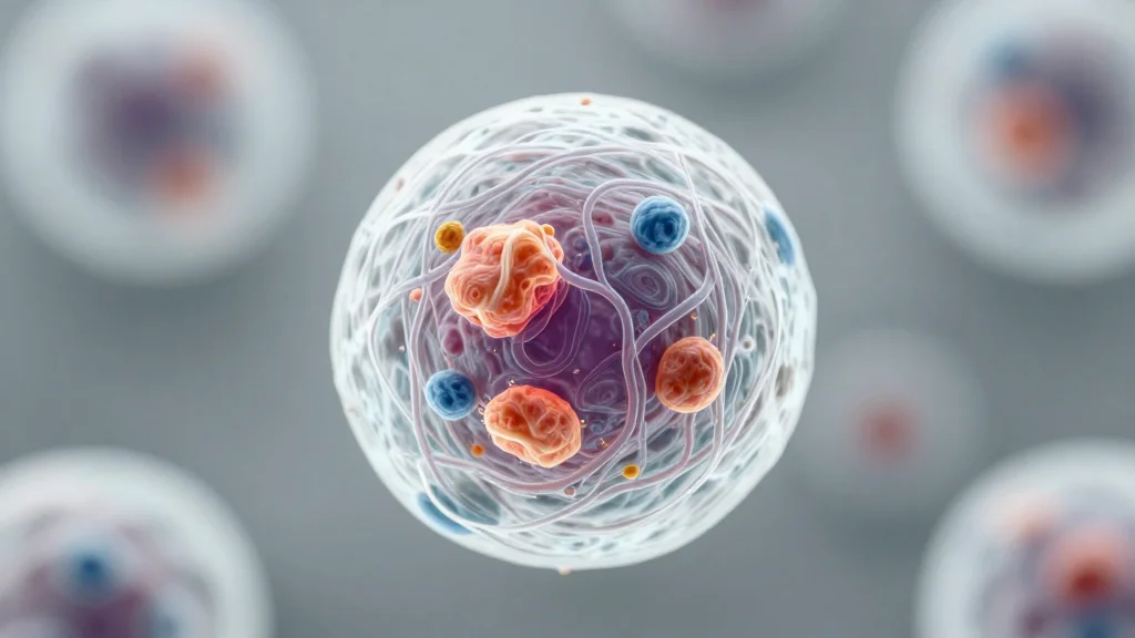
The ability to multiplex nanoscale imaging accelerates discovery of nuclear mechanisms underlying cancer and other diseases, paving the way for earlier diagnostics and targeted therapies.
Key Takeaways
- •DNA‑PAINT now tags 12 nuclear targets simultaneously
- •Achieves 3–5 nm resolution with faster, longer‑binding DNA tags
- •Images nine proteins in under four hours, cutting hours per target
- •Reveals protein re‑distribution during transcription inhibition in cancer cells
- •Potential to detect disease‑related nuclear changes before symptoms appear
Pulse Analysis
The latest iteration of DNA‑PAINT microscopy pushes the boundaries of cellular imaging by expanding its multiplexing capacity from a handful of markers to a dozen distinct targets. By engineering DNA tags that bind more rapidly and remain attached longer, the IISc team achieved a spatial resolution of just 3–5 nanometers—fine enough to distinguish individual protein complexes within the crowded nuclear environment. This technical leap also slashes acquisition time, enabling nine‑plex imaging in under four hours, a task that previously required days of continuous scanning.
Beyond the hardware improvements, the method offers a powerful lens on disease biology. When the researchers inhibited transcription in a cancer cell line, the high‑resolution maps revealed pronounced rearrangements of structural and transcriptional proteins, hinting at early‑stage nuclear remodeling that precedes overt phenotypic changes. Such insights could translate into biomarkers detectable before clinical symptoms emerge, providing a window for preemptive intervention. Moreover, the ability to observe multiple pathways simultaneously equips scientists to dissect how epigenetic regulators, repair factors, and chromatin remodelers coordinate in real time.
The broader biotech landscape stands to benefit as well. High‑throughput, nanometer‑scale imaging can accelerate drug discovery by allowing rapid assessment of how candidate compounds alter nuclear architecture or transcriptional hubs. It also dovetails with emerging spatial omics platforms, offering a complementary visual validation layer. As the technique matures and becomes more accessible, it may become a staple in both academic labs and pharmaceutical pipelines, driving a new era of precision cellular phenotyping.
High-resolution imaging shines light on nanoscale nuclear organization
Comments
Want to join the conversation?
Loading comments...