Mammalian Osteoderm Ultrastructure in the Armored Acomys Spiny Mouse Tail
Why It Matters
Understanding the spiny mouse’s natural armor provides insight into evolutionary convergence and offers a blueprint for bio‑inspired protective materials and regenerative medicine.
Key Takeaways
- •Osteoderms are calcium‑phosphate plates with bone‑like and tooth‑like structures
- •Plates follow an A‑B‑A pattern offset 26° from the tail axis
- •Aspect ratio averages 3.8; lacunar porosity about 4.5%
- •Distal third contains vascular channels; proximal third shows higher cellularity
- •Collagen fibers exhibit Sharpey's insertions and a hypomineralized surface band
Pulse Analysis
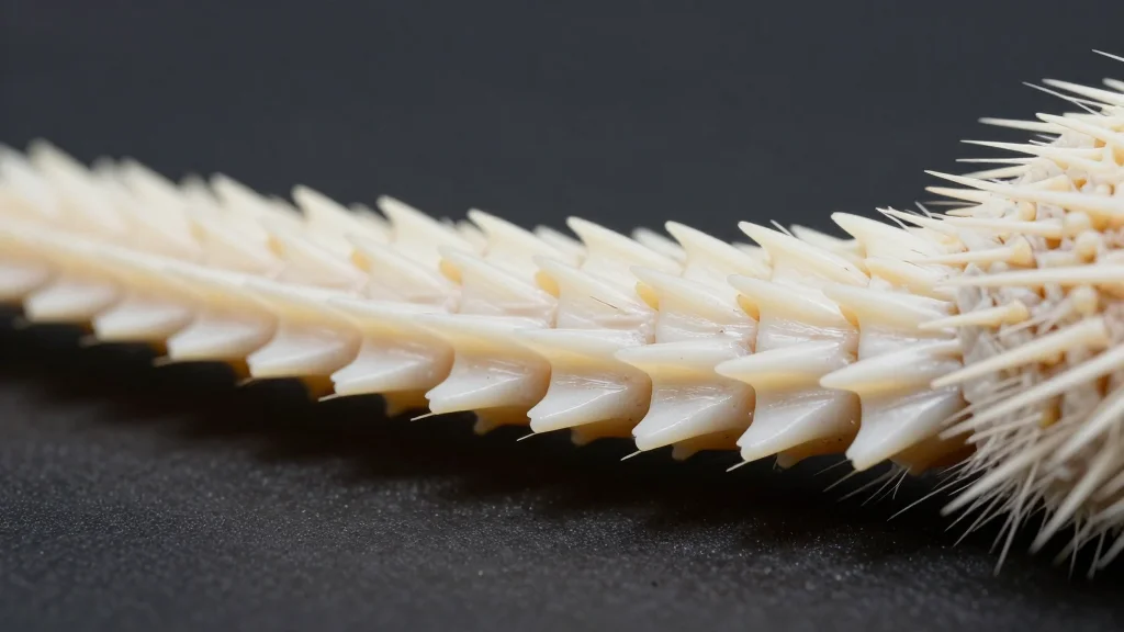
Mammalian armor is rare, but the spiny mouse (Acomys cahirinus) showcases a sophisticated version in its tail. Unlike the scutes of armadillos or reptilian osteoderms, these plates combine calcium‑phosphate mineralization with intricate collagen architecture. The convergence of bone‑like hardness and tooth‑like microstructures suggests a unique evolutionary solution to predation pressure, positioning the spiny mouse as a valuable model for comparative anatomy and functional morphology studies.
High‑resolution 2D and 3D microscopy revealed that each osteoderm aligns in a repeating A‑B sequence, tilted roughly 26° from the central tail axis. With an aspect ratio near 3.8 and a modest 4.5% lacunar porosity, the plates balance rigidity and flexibility. Vascular channels concentrate in the distal third, while the proximal region maintains slightly higher cellular density, indicating a growth gradient that may aid rapid regeneration after autotomy. Sharpey's fiber insertions anchor the dermal collagen to the mineral surface, creating a hypomineralized band that likely defines the fracture plane during tail shedding.
The implications extend beyond biology. Engineers seeking lightweight, impact‑resistant composites can draw inspiration from the spiny mouse’s hierarchical design, where mineral and organic phases cooperate to dissipate stress. In regenerative medicine, the patterned nucleation of mineral foci around hair follicles offers clues for guided bone growth and scaffold fabrication. As researchers decode the genetic and molecular pathways governing osteoderm formation, the spiny mouse may unlock new strategies for creating adaptive, self‑repairing materials that mimic nature’s own armor systems.
Mammalian Osteoderm Ultrastructure in the Armored Acomys Spiny Mouse Tail
Comments
Want to join the conversation?
Loading comments...