New Imaging Method Distinguishes Inflammation From Lung Fibrosis
Why It Matters
Identifying active inflammation non‑invasively enables clinicians to target anti‑inflammatory drugs to the right patients, improving outcomes and accelerating drug development in a high‑mortality disease area.
Key Takeaways
- •99mTc‑maraciclatide SPECT/CT distinguishes inflammation from fibrosis in ILD
- •Study involved 15 subjects: IPF, fibrotic HP, and healthy controls
- •Higher target‑to‑background ratios observed in ILD patients versus controls
- •FDA granted Fast Track status, aiming for market within two years
- •Phase 3 trial required before clinical adoption of the imaging method
Pulse Analysis
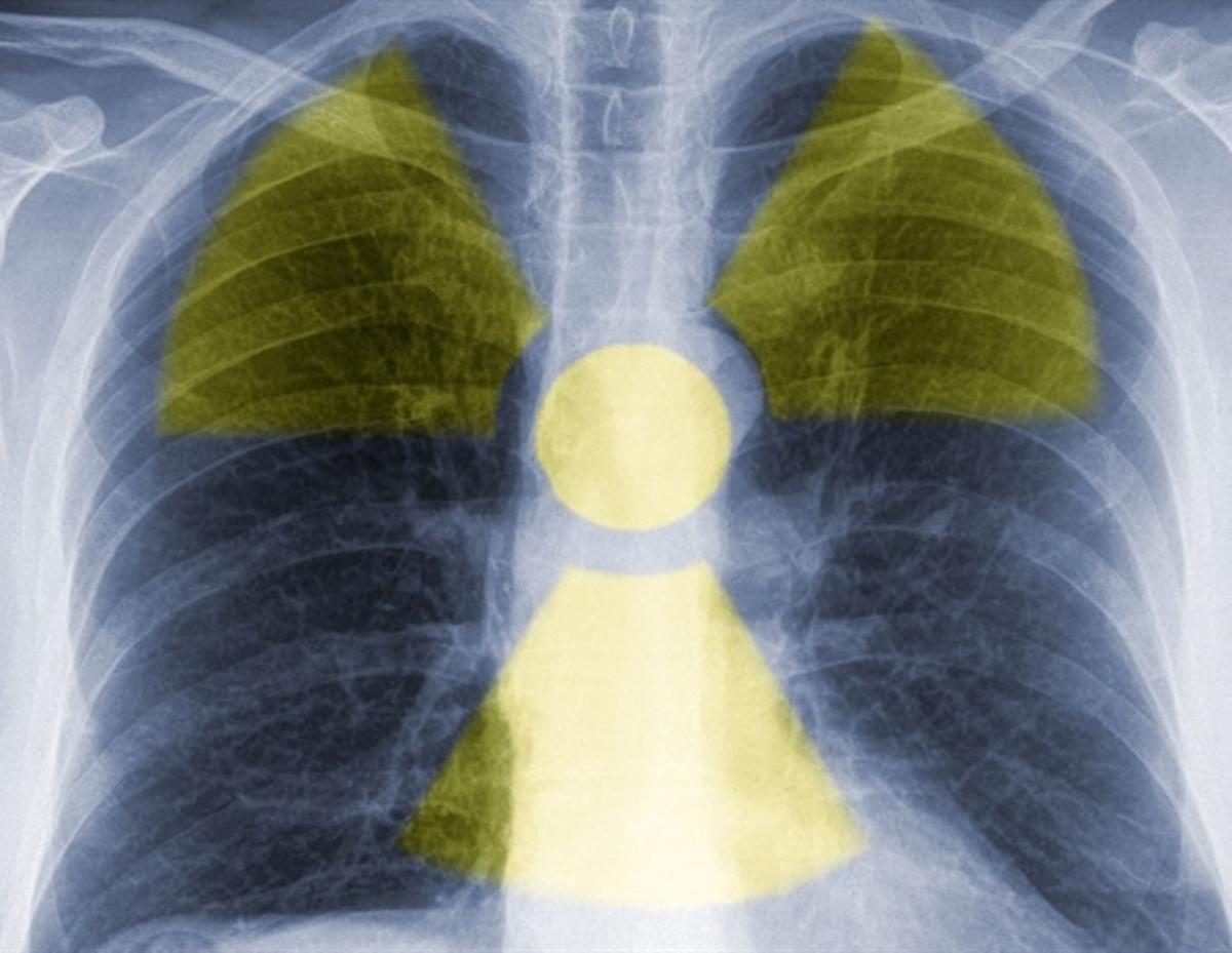
Interstitial lung disease (ILD) comprises more than 200 distinct disorders and affects roughly 650,000 Americans, accounting for up to 30,000 deaths each year. Clinicians have long struggled to separate active inflammation from irreversible fibrotic scarring, because conventional CT scans only reveal structural changes. Without a reliable, non‑invasive marker, patients may receive anti‑inflammatory drugs that offer little benefit and expose them to unnecessary side effects. The diagnostic gap has also slowed enrollment in trials of novel anti‑fibrotic and immunomodulatory agents.
The research presented at the 2026 SNMMI Annual Meeting introduced a 99mTc‑maraciclatide SPECT/CT protocol that visualizes angiogenesis, a hallmark of inflammation. In a pilot cohort of 15 participants—including five idiopathic pulmonary fibrosis patients, five fibrotic hypersensitivity pneumonitis cases, and five healthy volunteers—the tracer produced markedly higher target‑to‑background ratios in diseased lungs while remaining negligible in controls. FDA’s Fast Track designation reflects the agency’s confidence that the agent could fill a critical unmet need, and the sponsor anticipates a Phase 3 trial within the next 12‑18 months, potentially delivering a commercial product within two years.
From a commercial perspective, a validated inflammation‑specific imaging tool could reshape the ILD therapeutic landscape. Pharmaceutical firms developing anti‑inflammatory or anti‑fibrotic compounds would gain a companion diagnostic to stratify patients, improving trial success rates and enabling premium pricing for precision‑medicine regimens. Diagnostic imaging companies stand to add a high‑value nuclear‑medicine product to their portfolios, leveraging existing SPECT/CT infrastructure in hospitals worldwide. However, widespread adoption will hinge on reimbursement negotiations, demonstration of cost‑effectiveness, and the outcome of the forthcoming Phase 3 study.
New imaging method distinguishes inflammation from lung fibrosis
Comments
Want to join the conversation?
Loading comments...