Sea Squirt Reveals Glowing Spines and Unexpected Nervous System Anatomy
Why It Matters
The insights reshape our understanding of ascidian neuroanatomy and tunic biology, offering new comparative models for evolutionary and ecological research. Such knowledge could inform biomimetic material design and assessments of marine organism responses to environmental stressors.
Key Takeaways
- •Autofluorescent spines revealed in Halocynthia papillosa using multimodal imaging
- •Study maps spirally organized cellulose architecture of sea squirt tunic
- •Nervous system lacks typical cerebral ganglion thickening, indicating broader variation
- •3‑D tentacle reconstructions show species‑specific nerve and vessel patterns
- •Imaging platform enables future comparative studies of ascidian anatomy and ecology
Pulse Analysis
Ascidians, commonly called sea squirts, occupy a pivotal position at the junction of invertebrate and vertebrate evolution, making them a focal point for developmental and evolutionary biology. Traditional histology has struggled to resolve their low‑contrast tissues, but the advent of multimodal imaging—combining light microscopy, confocal fluorescence, magnetic resonance tomography, and high‑resolution synchrotron tomography—has opened a new window into their internal structures. By integrating these techniques, the team at Ruhr University Bochum generated true three‑dimensional reconstructions that capture both cellular detail and whole‑organ morphology, setting a benchmark for marine invertebrate imaging.
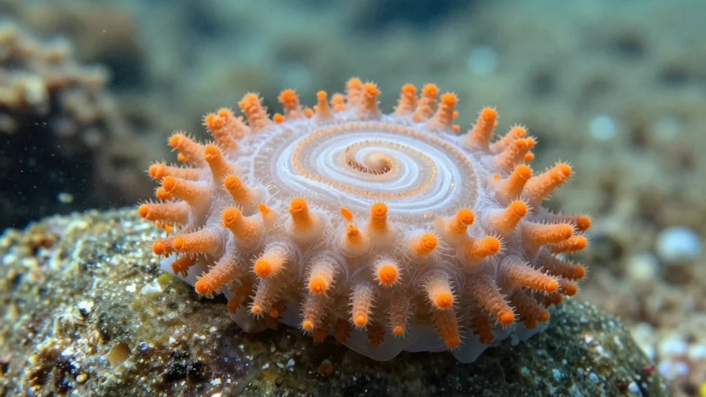
The investigation of Halocynthia papillosa uncovered a striking autofluorescent signal emanating from cuticular spines embedded in the tunic, a feature previously undocumented in adult ascidians. High‑resolution tomography revealed a spirally arranged cellulose lattice that likely contributes to the organism’s mechanical resilience. Equally surprising was the nervous system architecture: the expected cerebral ganglion thickening was absent, suggesting a broader spectrum of neural organization across ascidian species than recognized. Three‑dimensional models of the oral siphon tentacles further displayed species‑specific arrangements of nerves and blood vessels, highlighting functional specialization.
These discoveries have far‑reaching consequences for both basic science and applied research. A more nuanced map of ascidian neuroanatomy can refine phylogenetic models and illuminate the evolutionary steps leading to vertebrate central nervous systems. The unique cellulose architecture and fluorescent spines may inspire biomimetic materials with tunable optical or mechanical properties. Moreover, the imaging pipeline established by the researchers provides a scalable framework for assessing how environmental factors—such as underwater noise or temperature shifts—alter marine organism morphology. Future comparative studies across the 3,000‑plus ascidian species are now within practical reach.
Sea squirt reveals glowing spines and unexpected nervous system anatomy
Comments
Want to join the conversation?
Loading comments...