Spatial Proteomic Analysis in Human Alzheimer’s Disease Brains Enables Identification of Microenvironment-Dependent Microglial Cell States
Why It Matters
The study provides the first spatial proteomic map of human microglial diversity in Alzheimer’s, offering new mechanistic insights and potential therapeutic targets, while demonstrating that high‑parameter imaging can be applied to archived brain specimens.
Key Takeaways
- •CODEX-CNS adapts multiplex imaging for FFPE human brain tissue.
- •Study analyzed 51 samples, profiling 32 proteins at single‑cell resolution.
- •Identified microglial subpopulation enriched near dense Aβ plaques.
- •Morphology and protein clusters reveal microglia’s microenvironment‑dependent states.
- •Spatial proteomics uncovers cell‑cell interaction changes in AD versus controls.
Pulse Analysis
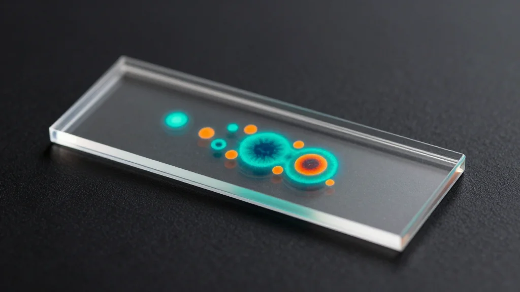
Understanding microglial heterogeneity in Alzheimer’s disease has been hampered by techniques that require tissue dissociation, losing spatial context. Traditional immunohistochemistry captures only a handful of markers, while single‑cell RNA sequencing lacks protein‑level detail and cannot map cells to pathological features such as amyloid plaques. By modifying the CODEX workflow to reduce autofluorescence and incorporate a brain‑specific segmentation pipeline, researchers now achieve simultaneous visualization of up to 32 proteins in archival FFPE sections. This breakthrough enables high‑resolution, spatially aware profiling of neurons, glia, vasculature, and disease hallmarks within the same tissue slice.
Applying CODEX‑CNS to frontal cortex specimens from 26 Alzheimer’s patients and 25 age‑matched controls uncovered five protein‑based myeloid clusters and three morphological microglial phenotypes. Notably, a microglial subpopulation co‑expressing CD163 and TMEM119 clustered tightly around dense Aβ plaques, suggesting a plaque‑responsive state distinct from classic disease‑associated microglia identified in mouse models. Morphological analysis further linked ramified microglia to dendritic markers, while rounded cells associated with vascular niches. Spatial interaction metrics revealed increased microglia‑microglia contacts and heightened astrocyte‑neuron proximity in diseased tissue, indicating altered cellular networks that may drive neurodegeneration.
These findings have immediate implications for Alzheimer’s research and drug development. By pinpointing microglial states that are defined by their immediate microenvironment, therapeutic strategies can be designed to modulate specific cell‑type interactions rather than broadly targeting inflammation. Moreover, the successful deployment of CODEX‑CNS on FFPE samples opens the door for large‑scale retrospective studies across brain banks, accelerating discovery of biomarkers and mechanistic pathways. As spatial proteomics matures, it is poised to become a cornerstone technology for decoding complex neurodegenerative processes in human tissue.
Spatial proteomic analysis in human Alzheimer’s disease brains enables identification of microenvironment-dependent microglial cell states
Comments
Want to join the conversation?
Loading comments...