Why It Matters
Understanding CPL assembly clarifies a key cause of early embryonic arrest, offering new targets for infertility treatment and maternal‑effect disease research.
Key Takeaways
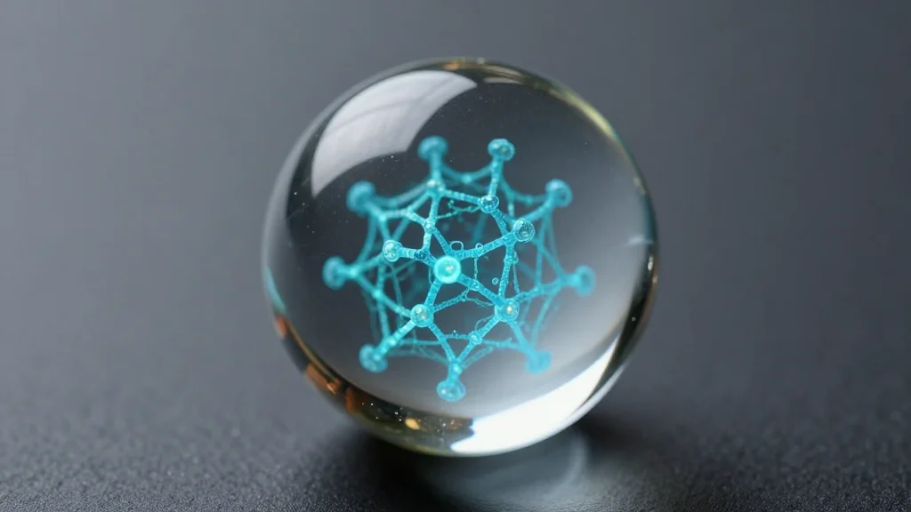
- •Cryo‑EM resolved 4 MDa CPL unit at 3.74 Å.
- •Framework built from PADI6 decamers and SCMC.
- •NLRP4F linkers polymerize frameworks into filaments.
- •Core sequesters UHRF1, tubulin heterodimers, inactive SCF complex.
- •Reveals CPLs as maternal proteostasis organelles for early development.
Pulse Analysis
The oocyte relies on a pre‑loaded inventory of proteins to jump‑start development before the embryonic genome awakens. Cytoplasmic lattices (CPLs) have long been recognized as storage hubs, yet their molecular organization remained speculative. By applying high‑resolution cryo‑EM to native mouse oocytes, the research team captured a 4‑megadalton repeating unit, exposing a three‑layered scaffold that integrates structural and regulatory components. This breakthrough bridges a critical knowledge gap, showing how maternal factors are physically sequestered and protected until fertilization triggers their release.
At the heart of the CPL, the study identifies a compact core where the epigenetic regulator UHRF1 is immobilized by PADI6, UBE2D, and NLRP14, preventing premature nuclear entry and ubiquitin ligase activity. Simultaneously, the core stocks GTP‑bound α/β‑tubulin heterodimers and an inactive SCF (FBXW‑SKP1) complex, effectively creating a ready‑to‑deploy microtubule and ubiquitination reservoir. The external framework, composed of PADI6 decamers and the subcortical maternal complex, is linked by polymerizing NLRP4F filaments, forming an extended filamentous network that can rapidly reorganize during the oocyte‑to‑embryo transition.
These insights have immediate implications for reproductive biology and medicine. Early embryonic arrest, a leading cause of infertility, often stems from disrupted CPL components; pinpointing the structural dependencies offers concrete molecular targets for diagnostics or therapeutic intervention. Moreover, the CPL model may inspire bio‑engineered nanostructures for controlled protein storage and release, extending its relevance beyond developmental biology into biotechnology and drug delivery. Future work will likely explore how CPL dynamics are regulated by post‑translational modifications and how similar organelles function in other species, deepening our grasp of maternal effect mechanisms.
Structure of the mouse cytoplasmic lattice
Comments
Want to join the conversation?
Loading comments...