3D Imaging Reveals Messy-Looking Supraparticles Can Be Nearly Perfect Crystals Inside
•March 10, 2026
0
Why It Matters
Understanding the hidden crystalline order enables precise design of photonic materials, accelerating their adoption in coatings and optical devices. The technique also offers a template for probing other colloidal systems where internal structure dictates performance.
Key Takeaways
- •3D microscopy reveals internal crystal order in supraparticles
- •Confocal + STED resolution improved fourfold over conventional methods
- •Machine learning classifies crystal structures from fluorescent particle coordinates
- •Potential for durable, angle‑independent color coatings in industry
Pulse Analysis
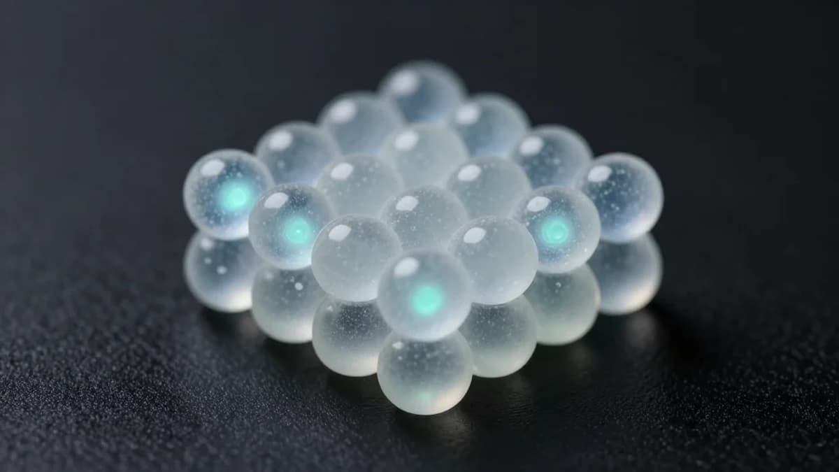
The breakthrough stems from marrying two cutting‑edge imaging modalities—confocal microscopy and STED (stimulated emission depletion) microscopy—to breach the diffraction limit that has long hampered nanoscale observation. By sharpening resolution roughly four times beyond standard confocal capabilities, the team could resolve sub‑500 nm silica particles in three dimensions, a scale comparable to visible wavelengths. This level of detail transforms what was once a surface‑only view into a volumetric map, exposing the true arrangement of thousands of colloids within each supraparticle.
Beyond hardware, the researchers embedded fluorescent cores into the supraparticles, allowing each constituent particle to be pinpointed in space. A bespoke machine‑learning model then parsed these coordinates, distinguishing subtle packing variations and assigning crystal classes automatically. This synergy of high‑resolution optics and AI reduces manual interpretation, accelerates data throughput, and sets a new standard for quantitative real‑space analysis in soft‑matter physics.
The implications extend far beyond academic curiosity. Photonic supraparticles mimic the angle‑independent coloration of Morpho butterflies, offering a pathway to pigments that retain hue without chemical dyes. Such materials could revolutionize paints, automotive finishes, and security inks by delivering long‑lasting, environmentally stable colour. Moreover, the methodology is broadly applicable to any colloidal system where internal order governs optical, mechanical, or catalytic properties, positioning it as a versatile tool for next‑generation material engineering.
3D imaging reveals messy-looking supraparticles can be nearly perfect crystals inside
0
Comments
Want to join the conversation?
Loading comments...