New SLAC Method Guides Better Cell Slice Preparation for Cryo-ET Imaging
•January 16, 2026
0
Why It Matters
The technique dramatically raises the efficiency and success rate of cryo‑ET, accelerating structural studies of rare or tiny cellular components and viral particles.
Key Takeaways
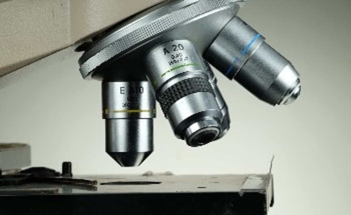
- •Tri‑coincident system aligns SEM, ion beam, and optical microscope.
- •Fluorescence interference signals locate targets during milling.
- •Milling accuracy improves roughly tenfold.
- •Enables cryo‑ET imaging of 26‑nm viruses.
- •Software automates dim‑bright pattern analysis for precision.
Pulse Analysis
Cryogenic electron tomography delivers near‑atomic 3‑D views of cellular interiors, but its power is limited by the need for ultra‑thin sections—typically under 200 nm—to let electrons pass. Conventional ion‑beam milling can produce such slices, yet researchers often waste time because they cannot verify whether the region of interest remains in the final lamella. This bottleneck hampers studies of low‑abundance structures, from ribosomal subunits to viral entry complexes, and slows the pipeline from sample to insight.
The SLAC team’s tri‑coincident platform solves the visibility problem by merging three instruments into a single, co‑aligned workspace. As the ion beam carves away the cell’s top layers, fluorescent tags on the target emit light that reflects off the freshly exposed surface, creating interference patterns that alternately dim and brighten. Custom software interprets these fluctuations in real time, delivering nanometer‑scale positional feedback. The result is a tenfold increase in milling precision, turning previously elusive targets—demonstrated with a 26‑nm virus—into routine cryo‑ET subjects.
Beyond immediate gains in throughput, this method opens new avenues for structural virology, drug discovery, and cell‑biology research that depend on visualizing transient or rare assemblies. By integrating advanced fluorescence modalities—such as super‑resolution or correlative light‑electron techniques—future iterations could push resolution limits further and automate target selection. Funding from DOE, NIH, and the Chan‑Zuckerberg Initiative underscores the broader scientific community’s appetite for tools that bridge optics and electron microscopy, positioning SLAC’s innovation as a catalyst for next‑generation cryo‑ET applications.
New SLAC Method Guides Better Cell Slice Preparation for Cryo-ET Imaging
0
Comments
Want to join the conversation?
Loading comments...