New Technique Maps Cancer Drug Uptake Inside Living Cells
Why It Matters
Accurate subcellular localization of radionuclide drugs accelerates development of therapies that maximize tumor kill while sparing healthy tissue, a critical hurdle for precision oncology. The approach also opens new research avenues for metal‑related disease mechanisms.
Key Takeaways
- •Method combines SEISMIC capillary sampling with LA‑ICP‑MS for subcellular metal detection
- •First live‑cell measurement of thallium distribution in pancreatic cancer cells
- •Enables tracking of radionuclide therapy localization, crucial for DNA‑targeted damage
- •Technique applicable to other metal‑based drugs and toxicants across diseases
- •Future work aims to sample nuclei and improve subcellular purity verification
Pulse Analysis
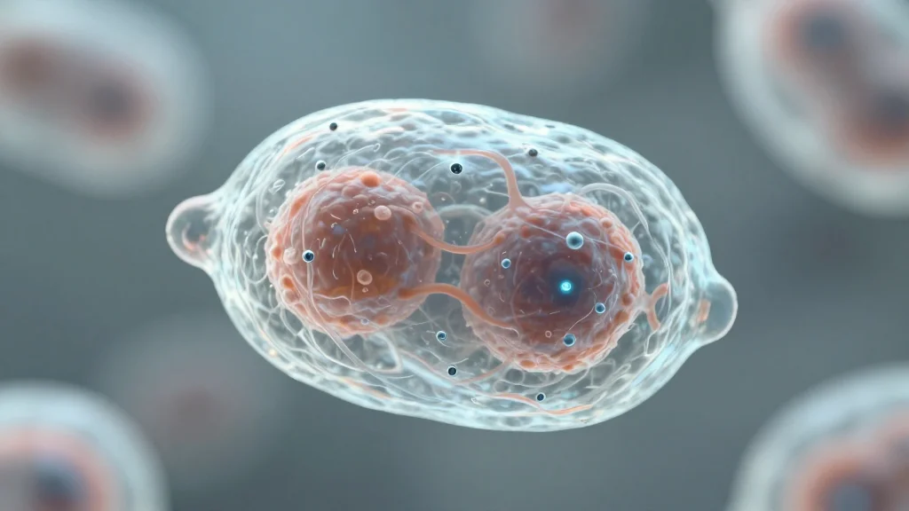
Targeted radionuclide therapy has emerged as a promising avenue for treating hard‑to‑reach tumors, delivering radiation directly to malignant cells while limiting exposure to surrounding tissue. Yet, the therapeutic promise hinges on a drug’s ability to reach the cell nucleus, where DNA damage triggers cell death. Conventional assays require cell fixation or lysis, obscuring real‑time distribution and potentially altering drug behavior. The lack of a live‑cell, subcellular mapping tool has long constrained researchers, slowing optimization of dose, carrier molecules, and delivery vectors.
The new workflow bridges that gap by integrating SEISMIC’s micromanipulation capillary sampling with laser‑ablation inductively coupled plasma mass spectrometry (LA‑ICP‑MS). Tiny glass tips extract whole cells or organelles—such as mitochondria—under a microscope, preserving cellular integrity. Subsequent laser ablation vaporizes the sampled material, allowing the ICP‑MS to quantify thallium at parts‑per‑trillion levels. In proof‑of‑concept experiments, thallium chloride, a stable analogue of the therapeutic thallium‑201 isotope, was detected inside individual pancreatic cancer cells and their mitochondria, confirming that the technique can resolve metal distribution at the organelle scale.
Beyond oncology, this capability reshapes how scientists investigate any metal‑based compound, from diagnostic agents to environmental toxins. By revealing precise intracellular locales, researchers can better correlate metal accumulation with functional outcomes, accelerating drug design and safety assessments. Ongoing refinements aim to sample nuclei directly and enhance purity checks, positioning the method as a versatile platform for precision medicine, toxicology, and metabolic research. As the field moves toward increasingly targeted therapeutics, tools that illuminate subcellular pathways will become indispensable for translating molecular insights into clinical breakthroughs.
New technique maps cancer drug uptake inside living cells
Comments
Want to join the conversation?
Loading comments...