Stanford Team Generates Light Inside Deep Tissue Using Ultrasound‑Activated Nanoparticles
Why It Matters
Non‑invasive light delivery has been a holy grail for both basic neuroscience and clinical medicine. Current optogenetic and photodynamic‑therapy workflows rely on implanted fiber‑optic probes, which limit the spatial reach of experiments and introduce infection risks. By using ultrasound—a modality already approved for diagnostic imaging—to activate nanophosphors, the Stanford work could democratize deep‑tissue illumination, making it accessible in outpatient settings and reducing procedural costs. Moreover, the platform illustrates how nanotechnology can turn ordinary physiological carriers (the bloodstream) into programmable light sources, opening a new design space for multifunctional therapeutic agents that combine sensing, imaging and actuation. Beyond the immediate biomedical promise, the technology showcases a broader trend in nanotech: leveraging external physical fields (acoustic, magnetic, electric) to control nanoscale functions inside the body. This paradigm reduces the need for permanent implants and aligns with regulatory preferences for reversible, on‑demand therapies. If the approach scales, it could accelerate the adoption of nanomaterial‑based interventions across oncology, cardiology and neurology, driving investment and competition in a field that has struggled to move from laboratory curiosity to market‑ready products.
Key Takeaways
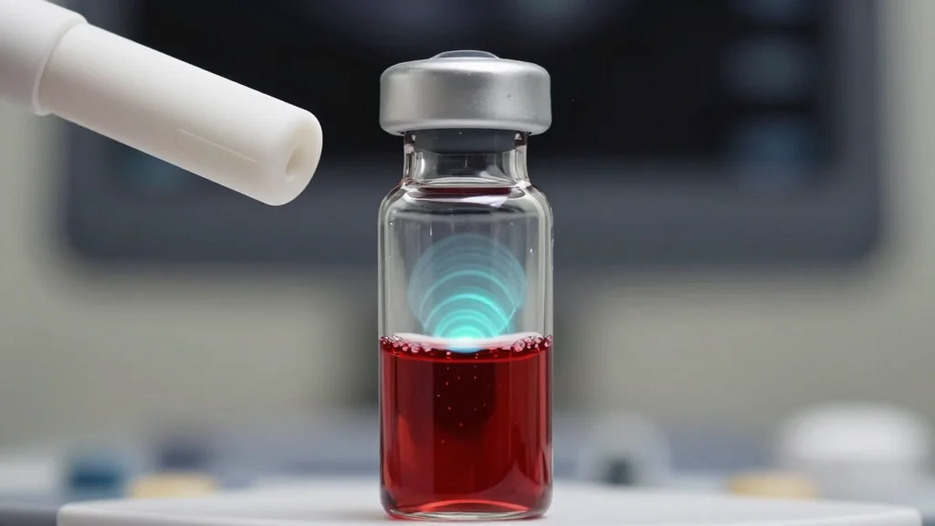
- •Stanford researchers demonstrated ultrasound‑triggered nanophosphors that emit blue light inside live mouse tissue.
- •Particles are ceramic nanocrystals coated with a biocompatible polymer, remaining dark until compressed by focused ultrasound.
- •Targeted illumination activated opsin‑expressing neurons, causing directional turning behavior in freely moving mice.
- •The system can scan the ultrasound focal point, creating multiple, movable light spots without any implanted hardware.
- •Future steps include scaling to larger animals, safety testing, and filing a provisional patent for clinical translation.
Pulse Analysis
The Stanford breakthrough arrives at a moment when the nanotech industry is searching for a clear, market‑ready application that can justify the high cost of material development and regulatory approval. Historically, nanomaterials have excelled in drug delivery but have struggled to demonstrate a unique therapeutic advantage beyond conventional formulations. By turning nanophosphors into on‑demand light sources, the team creates a functional capability that cannot be replicated by small‑molecule drugs or existing medical devices, potentially unlocking a new revenue stream for nanotech firms.
From a competitive standpoint, the approach pits acoustic‑based activation against the more established optical fiber and implantable LED technologies. While fiber optics offer precise, high‑intensity illumination, they are invasive and limited to superficial or surgically accessible sites. Ultrasound, by contrast, can reach deep organs without breaching the skin, but its spatial resolution is coarser and power delivery is constrained by tissue heating limits. The Stanford work narrows this gap by engineering nanoparticles that convert modest acoustic pressures into bright, localized emission, suggesting a viable middle ground where safety and efficacy can coexist.
Commercialization will hinge on three factors: scalability of nanoparticle synthesis, reproducibility of ultrasound targeting in heterogeneous human anatomy, and clear clinical endpoints that justify reimbursement. Companies that can mass‑produce the coated ceramic particles with tight size distribution will enjoy a cost advantage, while partnerships with ultrasound device manufacturers could accelerate regulatory pathways. If early trials demonstrate that the method can, for example, activate optogenetic circuits in patients with Parkinson's disease or deliver photodynamic therapy to pancreatic tumors, investors are likely to pour capital into a new class of “acoustic‑nanophotonic” platforms. The next 12‑18 months will therefore be critical as academic labs, startups and established med‑tech firms race to translate this proof‑of‑concept into a therapeutic reality.
Stanford Team Generates Light Inside Deep Tissue Using Ultrasound‑Activated Nanoparticles
Comments
Want to join the conversation?
Loading comments...